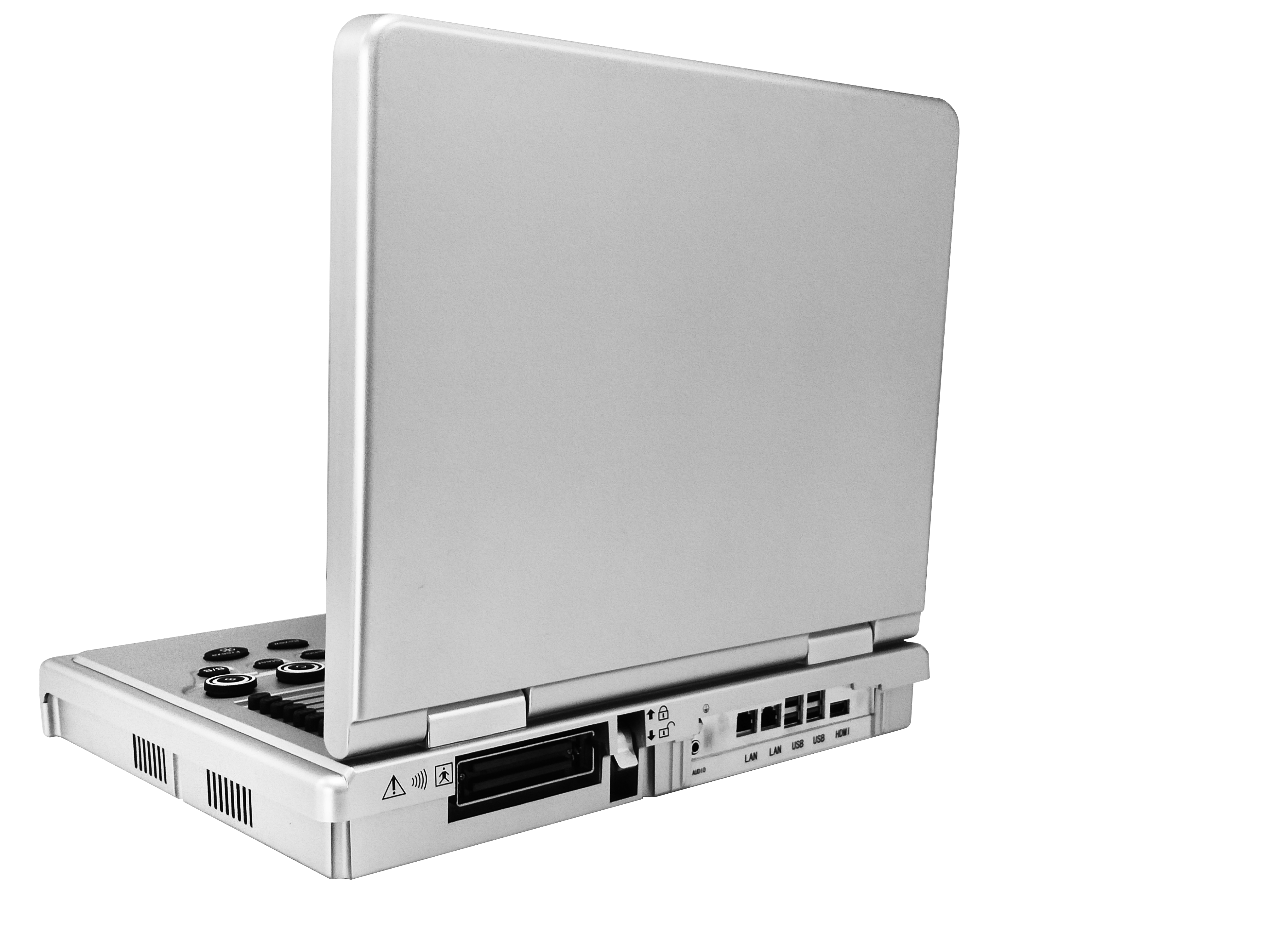
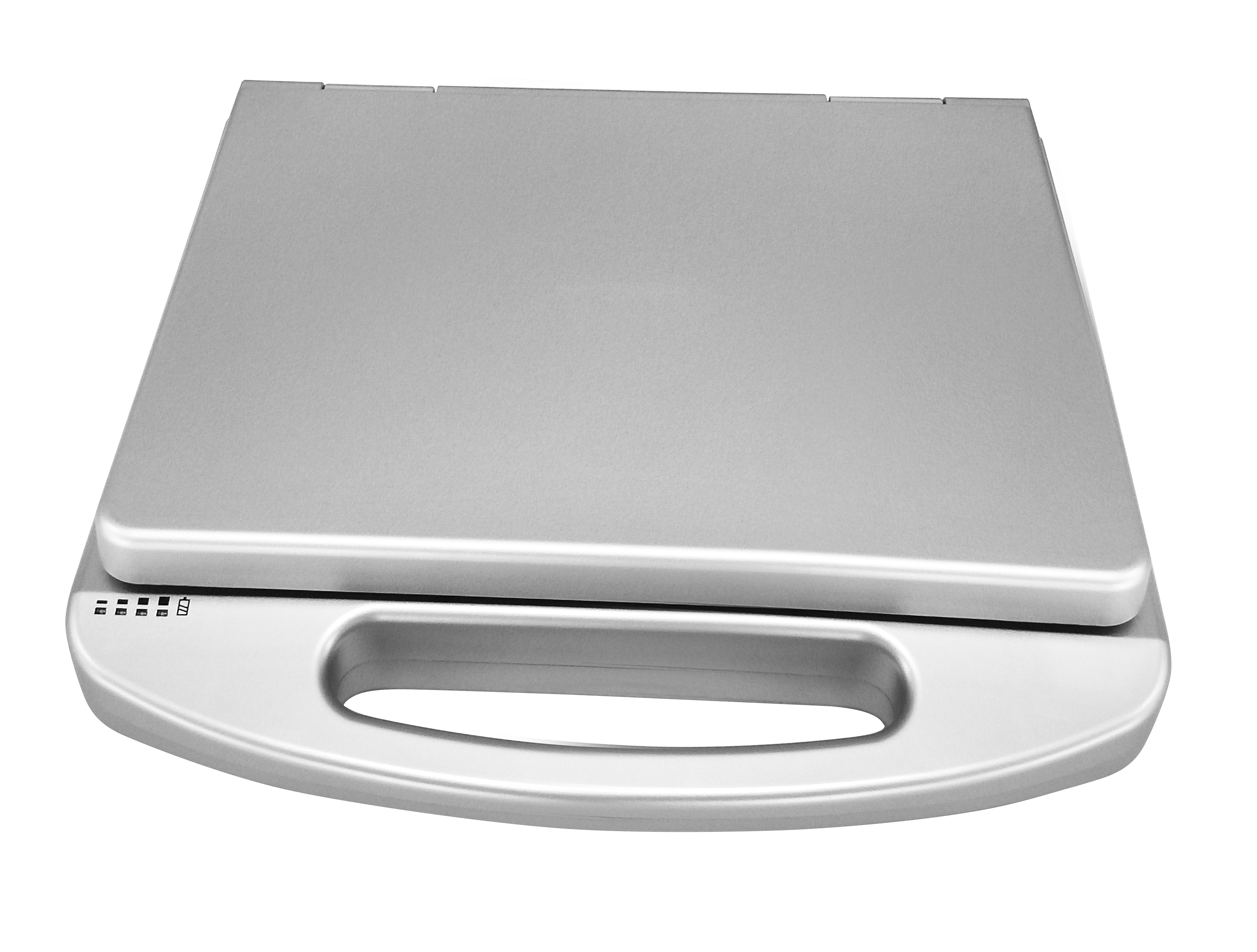
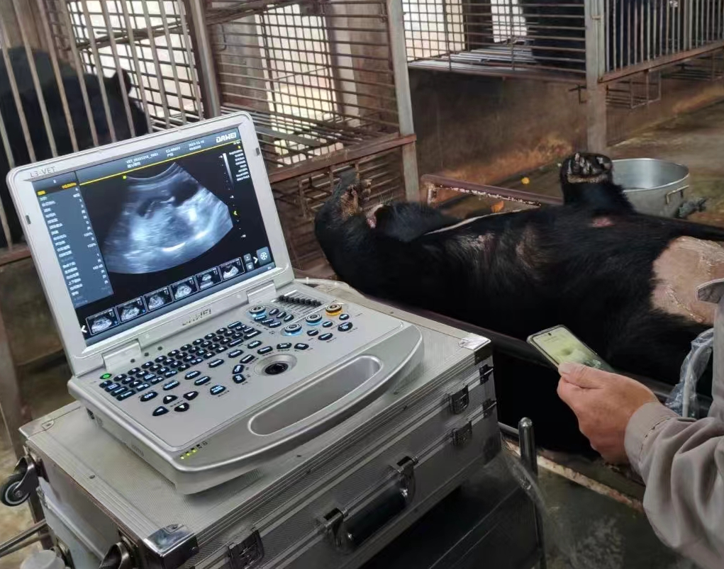
Main structure: Laptop type
Applications
Suitable for ultrasound examination for various needs in pet hospitals, clinics, zoos, breeding/breeding bases, and various research units.
Main Specifications And System Overview 1) Operating system: Windows 10 operating system
2) Spectral pulse Doppler
3) Directional energy Doppler
4) Real-time triplex
5) Spatial composite imaging function
6) Tissue harmonic imaging technology(THI)
7) 2B/4B imaging mode
8) System language options: Chinese, English, French, Russian, Spanish
9) Monitor: ≥ 15 inches
10) Integrated clipboard: shows saved images at the bottom of the screen, which can be directly dumped or deleted
11) The system has the function of on-the-spot upgrade.
12) Preset conditions: for different inspections, preset inspection conditions for optimizing images, reducing adjustments during operation, and external adjustments and combinations of adjustments required for common use
13) Probe interface ≥ 1
14) Trapezoidal imaging function
15) One-click intelligent optimization
Probes
1)Line array probe
2)Micro-convex probe (R11)
3)Micro-convex probe (R15)
4)Convex array probe
5)Rectal probe
Imaging Mode
1) Gain: 0-100, step 1 visually adjustable
2) TGC: 8 segments adjustable
3) Dynamic range: 20-280dB 20 levels visually adjustable
4) Pseudo-color: 0-11 levels, visually adjustable
5) Sound power: 5%-100%, in 5% steps, visually adjustable
6) Postural markers ≥ 18 kinds
7) Maximum number of focus points: 6 focus points, can be moved throughout
8) Grayscale: 0-7 levels of visual visibility adjustable
9) Filtering: 0-4
10) Scan range: 50%-100%
11) Frame correlation: 0-4 levels, visually adjustable
12) screen with Chinese form real-time display acoustic power, probe frequency, dynamic range, pseudo-color, grayscale and other 14 kinds of parameters can be adjusted
13) Scan line density: high school low
14) Noise Reduction: 0-14
Color Doppler Imaging Mode
1) Color frame correlation: 0-12 levels, visually adjustable
2) Color mapping: 0-7 levels, visually adjustable
3) Color flip: Adjustable
4) B/C split screen synchronous display function: with
5) Color baseline: 11 levels, visually adjustable
6) Color line density: high and low adjustable
7) Wall filtering: 0-5 levels adjustable
Spectral Doppler Imaging Mode 1) Sampling volume angle correction: -80°~80°adjustable
2) Sampling volume: 0.5mm-20mm visually adjustable
3) Frequency: 2.5MHz and 3.0Mhz etc. visually adjustable (different probe range)
4) Baseline: 11 levels adjustable
5) Pseudo-color spectrum: 0-5
6) display layout: ≥ 4 kinds of visual adjustable
7) Speed scale: 32.8-328cm / s (different probe range)
8) Spectrum envelope function: real-time automatic spectrum envelope, manual spectrum envelope, and other modes can be selected, and the system automatically analyzes and displays: PS, ED, PI, RI, S/D, HR, and other data
9) Grayscale: 0-7
10) Wall filtering: 0-8
11) Dynamic range: 10-90db step by step 5
12) Noise Reduction: 0-60
13) Volume: 0-100
Measurement And Analysis Functions
1) Measurement items include distance, area, angle, time, slope, heart rate, speed, acceleration, resistance index/beat index, and other specialized measurements
2) Having specialized measurement kits according to the different organs examined.
3) Measurement line color and line type can be adjusted (including activation color and completion color)
4) The position and font size of the measurement result display can be adjusted as required.
5) Specialized software packages: obstetrics, hepatobiliary, renal, bladder, cardiac, gastrointestinal, uterine, adrenal, pancreatic, vascular, etc.
Graphic Management System: picture saving format: BMP DCM JPG
1) host built-in ≥ 128G solid-state drive starts fast and stable
2) Movie playback: ≥ 600 frames
3) Built-in Chinese file information management system: can record the number ID, pet nickname, pet NO., owner's name, etc., and can search and manage by number ID, pet nickname, pet NO., owner's name, etc.
4) Report type ≥ 6, abdominal ultrasound report, cardiac ultrasound report, obstetric ultrasound report, small organ ultrasound report, urological ultrasound report, vascular ultrasound report, provide picture proof.
5) Quick report graphic management
6) With DICOM 3.0 protocol, can connect to PACS system
Interfaces
4 USB ports, 1 Audio, 1 HDMI, 2 RJ-45.
Configuration
1) Color Doppler ultrasound diagnostic system Mainframe 1 set
2) Probes: Micro-convex R11 probe (standard), line array probe (optional), rectal probe (optional), etc.
3) Video printer (optional), ultrasound medical cart (optional)


